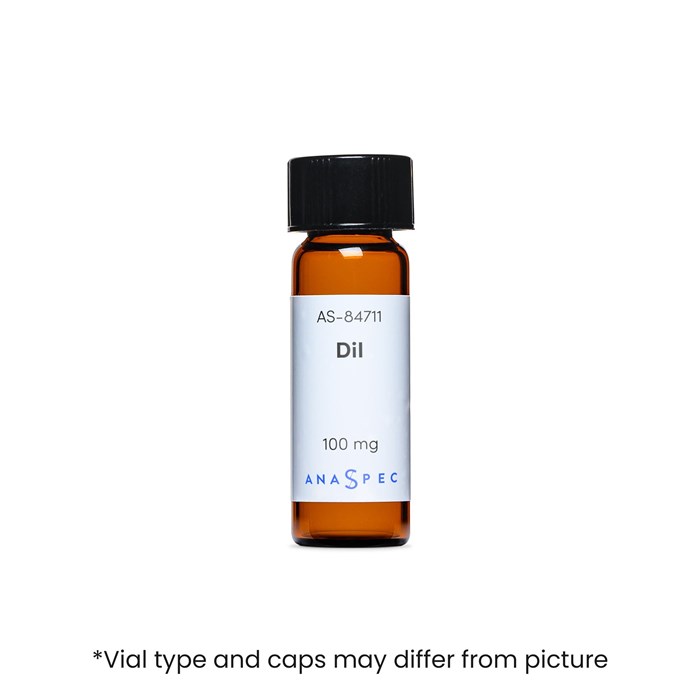
DiI [1,1'-Dioctadecyl-3,3,3',3'-tetramethylindocarbocyanine iodide] - 100 mg
- Cat.Number : AS-84711
- Manufacturer Ref. :
-
Availability :
In production
- Shipping conditions : Ice delivery fees must be applied
DiI, DiO, DiD and DiR dyes are a family of lipophilic fluorescent dyes which stain membranes and other lipophilic molecules. Their fluorescence highly increases when incorporated into membranes or bound to lipophilic molecules. Once applied onto cells, they diffuse laterally within the membranes to stain the entire cells. Their fluorescence is significantly increased upon membrane incorporation. This family of dyes fluoresces at distinct colors: DiI (orange), DiO (green), DiD (red) and DiR (far red); so they are adapted to multicolor imaging and flow cytometric of living cells. DiO and DiI can be used with standard FITC and TRITC filters respectively.
DiI is frequently used due to its very low cell toxicity. It is also used to label lipoproteins such as LDL and HDL.
Specifications
| Chemistry | |
| Molecular Formula |
|
|---|---|
| Molecular Mass/ Weight |
|
| Properties | |
| Absorbance (nm) |
|
| Emission (nm) |
|
| Color | |
| Storage & stability | |
| Form |
|
| Resuspension condition |
|
| Storage Conditions |
|
| Activity | |
| Application | |
| Detection Method | |
| Research Area | |
| Sub-category Research Area | |
| Usage |
|
| Codes | |
| Code Nacres |
|
You may also be interested in the following product(s)


Citations
Nuclear atrophy of retinal ganglion cells precedes the Bax-dependent stage of apoptosis.
Invest Ophthalmol Vis Sci . 2013 Mar 11 ; 54(3) 1805 | DOI : 10.1167/iovs.11-9310.
- K. Janssen
- et al
Evaluation of corticospinal axon loss by fluorescent dye tracing in mice with experimental autoimmune encephalomyelitis.
J Neurosci Methods . 2007 Aug 25 ; 167(2) 191 | DOI : 10.1016/j.jneumeth.2007.08.013
- Z. Liu
- et al
